HIGH TECHNOLOGY WHICH REWRITES THE RULES OF RADIOLOGY
HIRES SCAN
NewTom 5G allows to irradiate small portion of body, in order to see small anatomical details. This can be useful for proper implant assessment, because it requires the visualization of all aspects of the zygomatic bone, mandibular canal, maxillary sinus, tmj and other small parts, such as tooth roots, periodontal ligaments and any present pathology. Only 3D High Resolution imaging produces both the quality and the quantity of details necessary to accurately view those small details. Accuracy is one of the most important factors, together with dose reduction, also for analyzing middle and inner ear pathologies. NewTom has been recognized as an important diagnostic tool for this clinical fields: Oral and Maxillofacial Surgery, Implantology, TMJ, Oral Medicine, Orthodontics, Periodontology, Endodontics, Traumatology, ENT, Orthopedics and Veterinary.
PRECISE 1:1 SCALE IMAGING
With precise 1:1 scale imaging, NewTom technology eliminates the magnification errors of conventional cephalometric and opg imaging technology. 3D imaging allows the dental professional to identify potentially serious problems, such as airway passage obstructions and soft tissue abnormalities. CB3D imaging technology is the standard of care for implantologists, orthodontists, periodontists and oral/maxillofacial surgeons. Thanks to its design, more similar to a traditional CT, NewTom 5G helps the development of new fields of application.
ECOSCAN
The EcoScan is the novelty among the various scan protocols available on NewTom devices. This protocol reduces scan time and X-ray emission time as well as dosage, without affecting the high quality of the images.
SUPERIOR THIRD-PARTY COMPATIBILITY
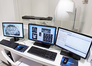
NewTom images are compatible with most major third-party software programs on the market as well as guided implant and maxillofacial surgery software. 3D imaging data is highly adaptable and can be imported and used in countless diagnostic and educational modes. Software segmentation adjusts the amount of soft tissue, underlines the hard tissue and accentuates the structure of the skull. Different intuitive software applications allow the creation of realistic models that can be positioned on images obtained from the scan. This creates infinite options that help in diagnosis, treatment planning, pre-surgical analysis, and patient education.
2D & 3D
It generates panoramics, cross sections and 3D bone models reading axial slices. This helps identifying all the anatomic aspects of the patient, the mandibular canal, the bone structure and the exact implant positions, in order to facilitate the surgery.
CB3D Versus MSCT
MSCT uses a narrow fan beam that rotates around the patient acquiring thin axial slices with each revolution. Cone Beam 3D imaging uses a cone-shaped beam to acquire the entire image in a scan using only one rotation.
SUPPORTED FORMATS
NewTom Implant Planning reads axial slices saved in DICOM 3.0 or in NNT format, which is the same format used by NewTom 5G, NewTom VGi, NewTom GiANO and previously released systems (NewTom VG, NewTom 3G and NewTom 9000).
BENEFITS
-A greater comfort for patients leads to a better acceptance of the treatment.
-SafeBeam™ Technology adjusts the radiation dosage for patient safety.
-Multiple FOV and different scan modes are selectable from the software and adaptable to various fields of application. In addition to boasting the smallest focal spot currently available on the market (0.3 mm), the unit also makes use of the revolutionary flat panel sensor technology, which provides for exceptional image quality and detail.
-The margin of error is reduced thanks to the precise 1:1 scale and a 16-bit grey scale.
-NNT software makes the image sharing process easier.
-Thanks to its revolutionary bed, the unit can be used to perform a wide range of examinations in various fields, such as dental, medical and veterinary.
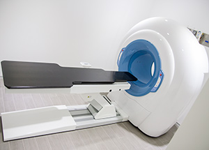
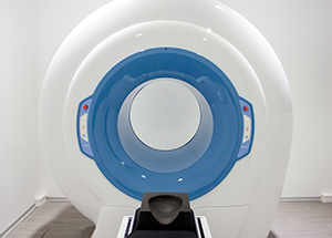
5G
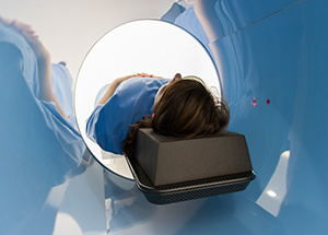
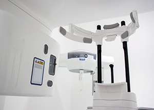
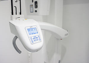
NewTom 5G, from the company that was the first to use the Cone Beam technology in dental field, represents the newest in CB3D technology. NewTom 5G takes an image at every degree of rotation, 360° rotation = 360 images, increasing the range of possibilities for image manipulation. It couples a revolutionary flat panel X-ray detector technology with a very small focal spot (0.3mm), to produce the clearest, sharpest images possible. NewTom 5G features an adjustable field of view, which allows doctors to irradiate just the right volume, depending on the different clinical applications. The size of FOV can vary from the smallest 6×6 cm to the biggest 18×16 cm and they can be selected directly from the software, before the scan. NewTom 5G emits a lower radiation in comparison with conventional CT, by using a “pulsed” emission, that unlike other systems, activates the X-ray source only when required and, for a full scan, it takes no more than 5 seconds of total exposure. With a fascinating design, users can explore new clinical applications, thanks to the open, pass-through style gantry and a motorized patient table. The supine position of the patient during the scan and the reduced scan time add comfort and stability for an excellent result in terms of image quality and patient satisfaction. The small footprint and the variable positioning make NewTom 5G the best choice for locations, where space is at a premium. NewTom 5G does not need air-conditioned rooms, its weight does not require reinforced floor and it can function in rooms without complicated and expensive radiation protection structures. All the operations executed by NewTom, the patient’s examination and the following calculations, are computer guided. The user, when performing the scan, is supported by user-friendly menus. Each step is associated to a mouse-activated icon. Following the same process, one can enter the integrated file of image-data.